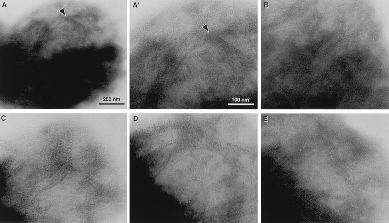
Figure 4.
Micrographs of intracellular filaments in detergent-treated, unfixed S. shibatae cells lightly stained with uranyl acetate, taken using intermediate voltage TEM (100–300 kV). A low (A) and high (A′) magnification of the same cell shows the distribution and distinct periodic structure of the intracellular filaments (arrowheads). Similar filaments were seen in many cells and four representative micrographs are shown (B–E).

