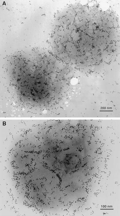
Figure 5.
ImmunoGold labeling of detergent-treated S. shibatae cells with polyclonal antibodies against the chaperonin seen at low (A) and high (B) magnification. The 5-nm gold particles (visible as black spots) distributed on the support grid around the cells suggests that chaperonins were released during sample preparation, but the abundance of gold particles remaining associated with cells indicates that a large number of chaperonins remained associated with an insoluble matrix. The pattern of gold particles in some regions is suggestive of filaments. Uranyl acetate-stained filaments, such as those shown in Fig. 4, were not seen in samples prepared for ImmunoGold labeling, therefore uranyl acetate staining was omitted to visualize better the distribution of gold particles.

