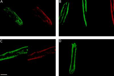
Figure 3.
TnI remodeling in adult single cardiac myocytes. Representative confocal images of indirect immunostaining of TnI in control (A and C) and AdCMVssTnI-infected (B and D) ventricular myocytes cultured for 4 (A and B) and 7 (C and D) days. TI-4 mAb labeling and cardiac specific TnI labeling with TI-1 mAb are shown in the left and right portions of A-D, respectively. (Bar = 17 μm.)

