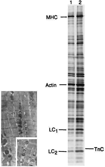
Figure 4.
(Right) Representative SDS/PAGE gel analysis of myofilament isoform composition and stoichiometry of AdCMVssTnI-treated (lane 1) and control (lane 2) ventricular myocytes. Gels indicate that isoform expression of myosin heavy chain, TnC, LC1, and LC2 are not altered by ssTnI gene transfer. Laser-based densitometry was performed, and the integrated peak for each protein was used to evaluate myofilament stoichiometry. Ratios for TnC/(TnC+LC1+LC2) were 0.06 and 0.08 in control and AdCMVssTnI-treated myocytes, respectively. Ratios for LC1 (LC1/(LC1+LC2) and LC2 (LC2/(LC1+LC2) were 0.56 and 0.45 in control myocytes and, 0.58 and 0.42 in AdCMVssTnI-treated myocytes, respectively. (Left) Transmission electron micrograph of a cardiac myocyte 3 days after adenovirus-mediated ssTnI gene transfer. The average sarcomere length of this cardiac myocyte is 1.7 μm. (Inset) Electron micrograph of a freshly isolated control cardiac myocyte with an average sarcomere length of 1.8 μm. Examination of other electron micrographs up to 5 days postinfection with AdCMVssTnI showed no differences in sarcomeric ultrastructure compared with control myocytes in primary culture. Ventricular myocytes in primary culture were fixed, embedded, and mounted as described previously (3).

