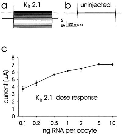
Figure 2.
(a and b) Typical two-electrode voltage clamp records (a) from an oocyte injected with Kir2.1 and (b) from an uninjected oocyte. The holding potential was −60 mV with 350 msec voltage steps to between 100 mV and −140 mV in −10 mV increments. Currents recorded at different step potentials are superimposed. The dotted lines indicate zero current level. (c) Inward current magnitudes recorded at −80 mV from oocytes injected with a range of concentrations of Kir2.1 cRNA. Data points represent mean ± SEM from five oocytes each, after subtraction of the mean current (inward 0.40 μA) recorded from five uninjected oocytes (SEM = 0.05 μA). Inward currents are presented as positive values for ease of presentation of the dose response.

