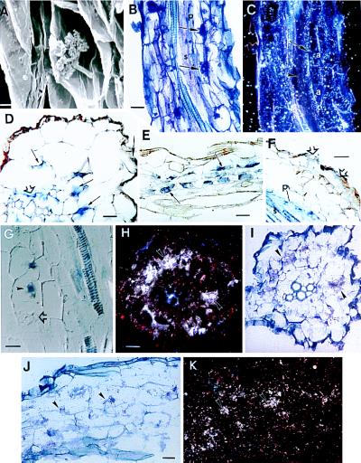
Figure 3.
(A) Scanning electron micrograph of an arbuscule in an alfalfa root. (Bar = 5 μm.) (B) Bright-field micrograph of a mycorrhizal root sectioned longitudinally. Arrows indicate arbuscules. The pericycle (p) and stele (s) are indicated. (Bar = 22 μm.) (C) Dark-field micrograph of B illustrating the presence of MsENOD40 transcripts (shown here as white dots representing silver grains) in root cortical cells surround the arbuscules (a) and in the pericycle (arrow), but not in the central part of the stele (arrowhead); 35S-labeled probe. Exposed for 6 weeks. (D) Bright-field micrograph of a transverse section of a young mycorrhizal root; arbuscules are not fully developed. Blue color indicating the presence of MsENOD40 transcript is detected in the pericycle (open arrow), the inner cortical cells (small arrows), and an epidermal cell (arrow); DIG-labeled probe. (Bar = 22 μm.) (E) Off-median longitudinal section through the inner cortex of a root. Arbuscules (small arrows) are in the inner cortical cells, and the blue color indicating the presence of MsENOD40 mRNAs is present in these cells; DIG-labeled probe. (Bar = 44 μm.) (F) Longitudinal section. Blue color is found in the epidermal cells (open arrows) as well as the pericycle (p) and stele (s); DIG-labeled probe. (Bar = 22 μm.) (G) Longitudinal section of a mycorrhizal root taken with Nomarski optics. Blue color indicating MsENOD40 transcript localization is found in the stele and pericycle and in two infected cells (arrowhead). Note that the fully developed arbuscule (open arrow) does not show the blue color; DIG-labeled probe. (Bar = 22 μm.) (H) Dark-field micrograph of a transverse section of mycorrhizal root. Silver grains indicating MsENOD2 mRNAs are clustered over the inner cortical cells, which contain arbuscules (arrows); 35S-labeled probe. Exposed for 3 weeks. (Bar = 22 μm.) (I) Bright-field micrograph of H. Arbuscules (stained purple, arrowheads) are within inner cortical cells. (J) Bright-field micrograph of an off-median longitudinally sectioned mycorrhizal root. The arrowheads indicate the arbuscules. (Bar = 22 μm.) (K) Dark-field view of J. Silver grains indicating MsENOD2 mRNAs are clustered over the arbuscules (arrows); 35S-labeled probe. Exposed for 3 weeks.

