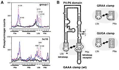
Figure 5.
Folding of the P4–P6 domain. (A) Dimethyl sulfate (DMS) modification patterns for the L5b tetraloop region within either the Tetrahymena group I or hc16 ligase ribozyme, based on primer extension analysis using reverse transcriptase (27). Thick line, no DMS and 50 mM MgCl2; thin line, DMS and 50 mM MgCl2; dashed line, DMS and 1 mM Na2EDTA. Nucleotide numbering follows the sequence of each ribozyme. After renaturation, the RNA (0.3 μM) was preincubated at 30°C for 5 min in 40 μl containing 200 mM KCl, 5 mM spermidine, 50 mM cacodylate (pH 7.5), and either MgCl2 or Na2EDTA, followed by addition of 1 μl of DMS in ethanol to a final concentration of 17 mM and incubation at 30°C for 5 min. (B) Tertiary interaction between the GAAA tetraloop of L5b and the corresponding tetraloop receptor of P6a (Left). Analogous interactions occur with the GRAA and GUGA clamps (Right).

