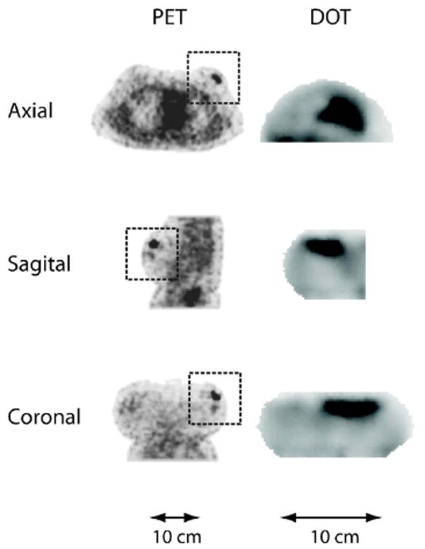
Fig. 3.

Axial, sagittal, and coronal slices from the PET (FDG) and DOT (μeff) reconstructed images of subject 4. The orientation for the PET and DOT images is the same. However, the DOT images are of the left breast only, whereas the entire torso is shown in the PET images. Rectangular boxes denote the breast region in the whole-body PET images.
