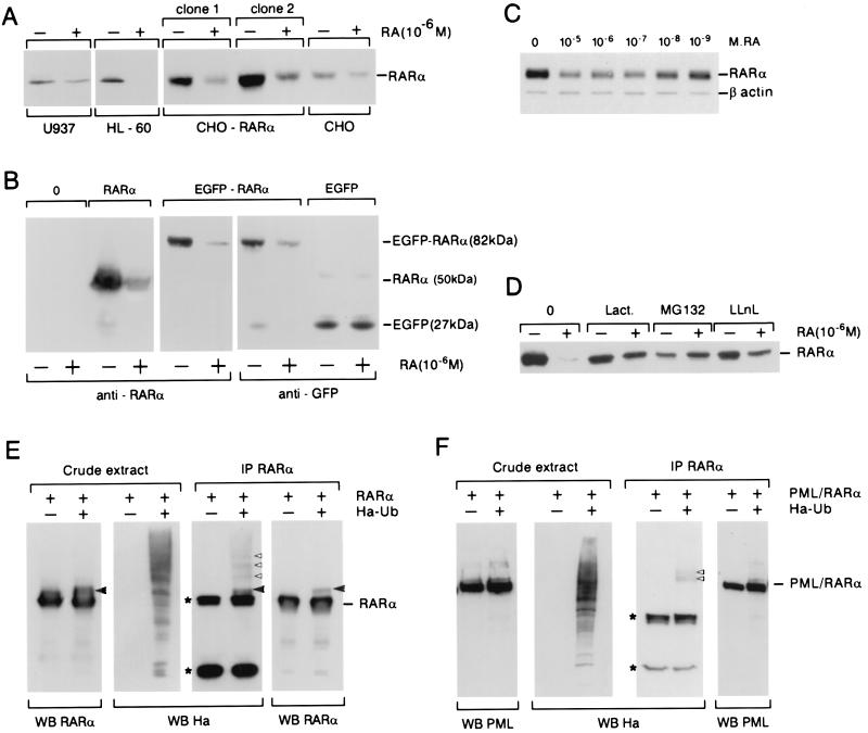
Figure 2.
Endogenous or transfected RARα is catabolized after RA exposure. (A) RARα is down-regulated after an overnight exposure to 10−6 M RA in U937 and HL-60 cells as well as in parental CHO cells or clones overexpressing RARα1 from a retroviral vector. (B) COS cells were transfected with expression vectors encoding RARα, EGFP-RARα, or EGFP and were or were not treated with RA overnight. Western blot analysis was performed with anti-RARα antibodies (Left) or anti-GFP antibodies (Right). (C) COS cells were transfected with RARα, translation was blocked with CHX, and various doses of RA applied for 5 h as indicated. An actin control is provided. (D) Proteasome inhibitors (10−6 M) block RA-induced RARα catabolism. (E and F) RARα or PML/RARα degradation is associated to receptor ubiquitination. COS cells were cotransfected with expression vectors encoding RARα or PML/RARα and (or and not) Ha-tagged ubiquitin. Cells exposed to RA for 3 h either were or were not immunoprecipitated (IP). Western blot analysis was performed by using anti-RARα, anti-PML, or anti-Ha as indicated. *, Heavy and light chains of the mouse antibodies used for immunoprecipitation; ◂, a RARα–ubiquitin adduct observed with both anti-Ha and anti-RAR antibodies, whereas those marked by ▹ are visible with anti-Ha alone.

