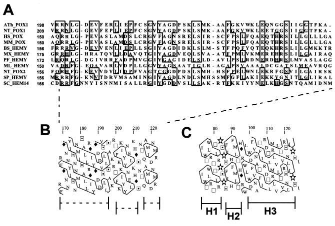
Figure 1.
(A) Sequence alignment of the putative membrane-anchoring domain in protoporphyrinogen oxidases (clustal w). (B) Two-dimensional graph representation [Hydrophobic Cluster Analysis (13)] of the peptide sequence of yeast protoporphyrinogen oxidase putative membrane-anchoring domain. (C) Two-dimensional graph representation (Hydrophobic Cluster Analysis) of the peptide sequence of sheep prostaglandin H2-synthase-1 membrane-anchoring domain determined from the crystal structure of the protein (11). H1, H2, and H3 are the amphipathic helices in interaction with the lipid bilayer.

