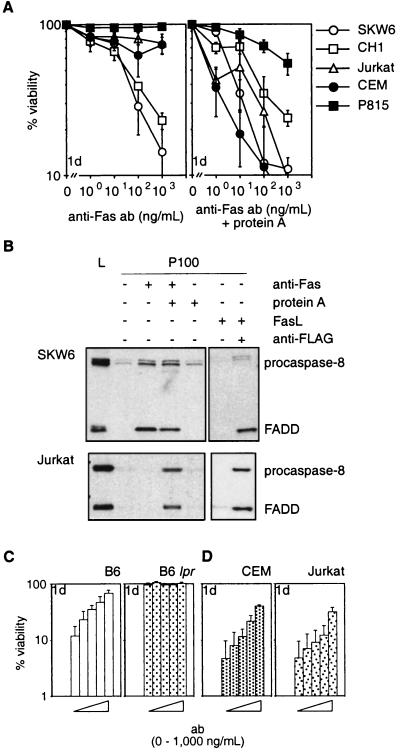
Figure 2.
Anti-Fas antibodies do not reliably mimic FasL. (A) SKW6, CH1, Jurkat, CEM, and P815 cells were treated with 0–103 ng/ml anti-Fas mAbs (APO-1 for human or Jo2 for mouse cells) without (Left) or with (Right) 100 ng/ml protein A. (B) Recruitment of FADD and procaspase-8 to the plasma membrane in SKW6 (Upper) and Jurkat (Lower) cells. Gels were loaded with total lysate (L) or equivalent pellet fractions (P100) from untreated cells or cells treated with anti-Fas mAb (APO-1) ± protein A or recombinant FasL ± anti-FLAG. (C) Purified mouse T cells from C57BL/6 (Left) or C57BL/6 lpr mice (Right) were incubated for 2 hr with Jo2 anti-mouse Fas mAb (bars from left to right: 0, 1, 10, 100, or 1,000 ng/ml) and then cocultured with Neuro2A-FasL or control cells. (D) After preincubation with APO-1 mAb (bars from left to right: 0, 1, 10, 100, or 1,000 ng/ml), CEM (Left), or Jurkat cells (Right) were cocultured with Neuro2A-FasL or control cells. Viability of cells cocultured with Neuro2A-FasL cells relative to cells cultured with control Neuro2A cells was determined after 1 day. Data shown represent arithmetic means ± SD of three independent experiments.

