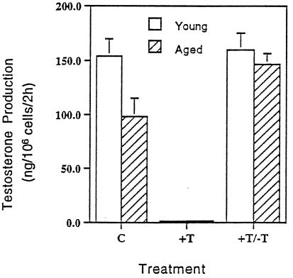
Figure 1.
The capacity of Leydig cells to produce testosterone, assayed in vitro under maximally stimulating LH. Within each experimental group of eight rats, there were three subgroups of 2–3 rats from which Leydig cells were isolated and pooled. The cells were run in triplicate. The graph shows the mean of the three subgroups ± SEM. C, Control rats of 3 and 13 months of age received empty implants for 8 months; the implants were removed; and 2 months later, when the rats were 13 (young) and 23 (aged) months of age, respectively, Leydig cells were isolated and assessed for their ability to produce testosterone. Leydig cells from the 23-month-old control rats produced significantly less testosterone than cells from the 13-month-old controls. +T, Leydig cells isolated from 11- and 21-month-old rats that had received exogenous testosterone for 8 months produced little testosterone. +T/−T, Two months after the implants were removed, when the rats were 13 and 23 months of age, respectively, the ability of the Leydig cells to produce testosterone was equivalent, in both cases at the high level of young controls.

