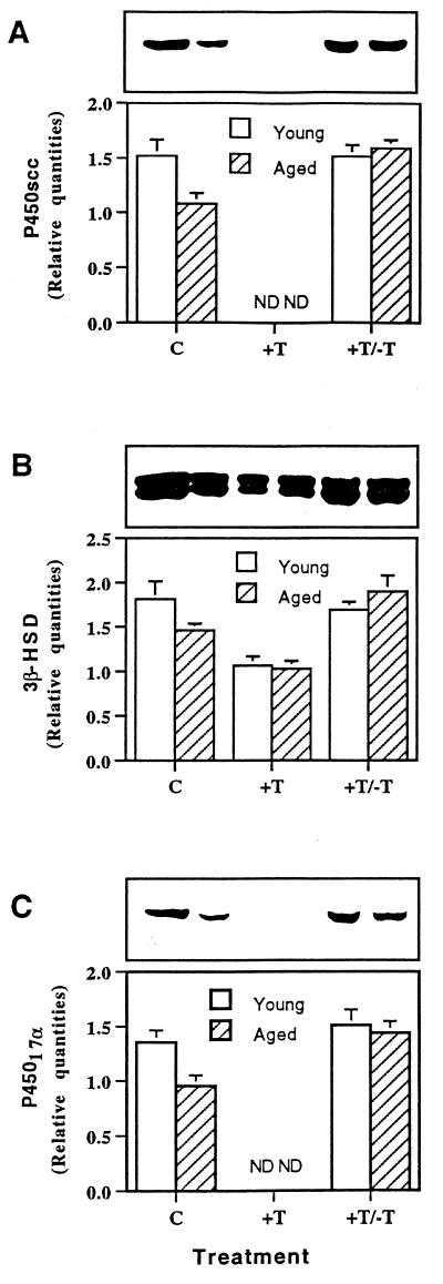
Figure 3.
Analysis of Leydig cell steroidogenic enzyme proteins by Western blot. Above each graph is a representative Western blot. Proteins (P450scc, 3β-HSD, and P45017a) were quantified by densitometric scanning of blots from three different pools of cells. C, Control rats of 3 and 13 months of age received empty implants for 8 months, the implants were removed, and 2 months later, when the rats were 13 and 23 months of age, respectively, Leydig cells were isolated and protein from equal numbers of cells was analyzed. The quantities of the P450scc (A), 3β-HSD (B), and P45017a (C) were reduced in cells of the 23-month-old compared with the 13-month-old rats. +T, After 8 months of testosterone treatment, all three enzymes were reduced, in the case of P450scc and P45017a, to undetectable levels. +T/−T, Two months after the removal of the implants, when the rats were 13 and 23 months of age, respectively, the steroidogenic enzyme levels in both age groups were restored to those comparable to the 13-month-old controls, significantly higher than the 23-month-old controls. Means ± SEM are shown. ND, not detectable.

