Figure 2.
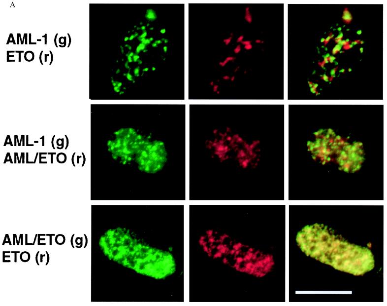
AML1B and AML/ETO proteins are targeted to distinct subnuclear locations. Immunofluorescence localization of coexpressed proteins in Saos-2 cells was examined in in situ nuclear-matrix preparations by using digital fluorescence microscopy (A) or laser-scanning confocal microscopy (B). AML and ETO coexpression (Top), AML and AML/ETO coexpression (Middle), and AML/ETO and ETO coexpression (Bottom). Epitope-tagged proteins were detected with specific antibodies and visualized by using a Texas red-conjugated secondary antibody seen as a red fluorescent signal (r, red, Center), and either a FITC secondary antibody or an EGFP epitope tag, seen as a green fluorescent signal (g, green, Left). Colocalization of red and green immunofluorescence signals is observed as yellow staining in the merged images (Right).


