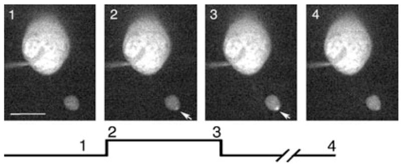
Fig. 5.

Addition of ryanodine (25 μM) to the pipette solution reduced the secondary spread of calcium through the rod terminal. Changes in [Ca2+]i in a single confocal slice from a rod terminal in the retinal slice evoked by depolarizing steps (−70 to −10 mV, 500 ms). The images show the terminal and cell soma. Image timing is shown diagramatically below the images. Image 1 shows a control image obtained prior to the step. Image 2 shows the first image obtained during the test step. Image 3 shows the last image obtained during the 500-ms test step. Arrows indicate a local hot spot of Ca2+ increase. Pipette solution contained the low-affinity Ca2+ sensitive dye Oregon Green 488 BAPTA-5N (100 μM). Image acquisition time: 48 ms. Scale bar, 10 μm.
