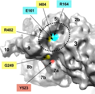
Fig. 5.
Identification of MH/CCD-linked residues in the FKBP12 binding pocket. The surface of the clamp in RyR1 is color-coded based on location of residues associated with MH and CCD mutations in the N-terminal homology models docked to the 3D map of RyR1. Cyan surface corresponds to residues from model 1, and yellow and red correspond to residues from model 2 (see color code used in Figs. 3 and 4). Putative surface-exposed mutation residues are labeled. The basin between subregions 3, 5, and 9 (indicated with dashed line) is proposed to form the FKBP-12 binding site in RyR1 (31, 32) and is identified to include four surface-exposed MH/CCD mutation sites (E161, R164, R402, and I404). The surface of the adjacent RyR1 subunit is shown with a mesh.

