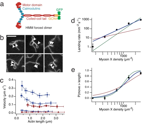
Fig. 1.
Motility of myosin X on single actin filaments. (a) Cartoon of the myosin X construct used in this study. The forced dimer was used for all in vitro assays. (b) Images of actin filament plectonemes in the gliding filament assay. Arrowheads indicate the extent of supercoiled segments. (Scale bar, 5 μm.) (c) Myosin X velocity depends on actin filament length. Measured actin filaments were binned by length, and velocities of myosin X (blue) and myosin VI (red) are shown (n = 10 filaments per point, ±SD in velocity and length). ATP concentration; 2 mM (circles), 1 mM (squares), and 1 μM (triangles). (d and e) Actin filament landing rate (d) and fraction of filaments that move greater than their length (e), both as a function of myosin X density on the surface. Shown are fits to models where one (red), two (blue), and three (green) motors are required to propel actin. Best fits are obtained for the two motor model: reduced χ2 = 3.7 (d), reduced χ2 = 1.4 (e). Error bars are standard errors obtained from counting statistics.

