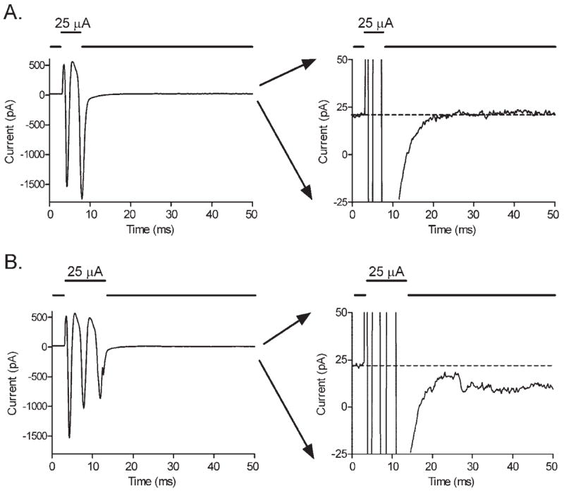
Figure 3.

(A) Pulses of extracellular current stimulation (5 milliseconds, 25 μA) evoked transient inward currents arising from action potentials in retinal ganglion cells. No sustained inward currents are seen (the trace in the right panel returned rapidly to baseline). (B) Longer pulses (10 milliseconds, 25 μA) evoked the same transient currents followed by small, sustained inward currents (the trace in the right panel did not return fully to baseline). The ordinate is expanded in panels at the right to better illustrate the small, sustained inward current evoked by 10-millisecond, but not 5-millisecond, steps.
