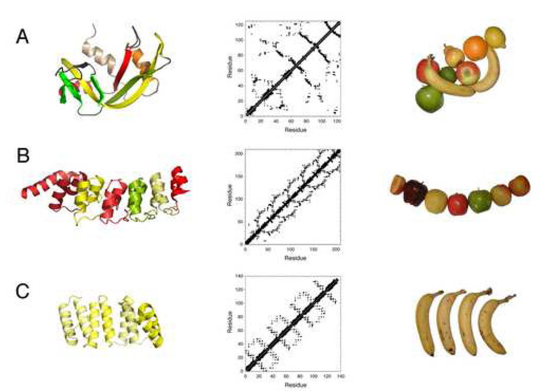
Figure 1. Comparison of globular and repeat protein structures.

(A) A typical globular protein used in folding studies (RNase A, top; 7RSA.pdb) is compared to (B) a naturally occurring repeat protein (the Notch ankyrin domain, middle; 1OT8.pdb chain A) and to (C) a consensus repeat protein (bottom; 2FO7.pdb). Left: ribbon diagrams (prepared with MacPyMOL [107]) coloring different secondary structure elements (top) and repeats (center, bottom). Middle: contact maps, emphasizing the lack of long-range contacts in repeat proteins (center, bottom), and regular patterns of tertiary structure in different regions of repeat proteins. Right: analogy of globular and repeat proteins to assemblages of fresh fruit. Although the usual fruits of the familiar metaphorical comparison are apples and oranges, some elements of secondary structure (and whole repeat units) are elongated and can be better represented by bananas.
