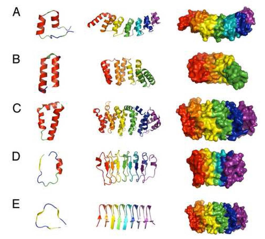
Figure 2. Structure of various repeat proteins.

(A) the ankyrin repeats of the Notch receptor, 1O8T.pdb; (B) consensus tetratricorepeats, 2F07.pdb; (C) heat repeats; 1UPK.pdb; (D) internalin-B leucine-rich repeats, 1H62.pdb; (E) hexapeptide repeats, 1J2Z.pdb. Left: overall architecture of single repeats of some of the most common linear repeat proteins. α-helices are red, β-strands are yellow, PPII structure is green, and tight turns are blue. Center: linear arrays of these the same repeats, with adjacent repeats colored from red to purple (N to C). Right: surface representation of adjacent repeats, showing contiguous packing over the entire domain.
