Fig. 1.
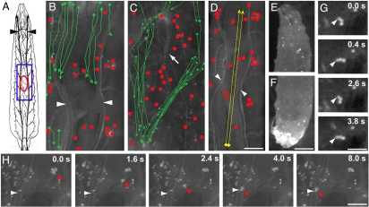
Circulating and tissue-bound blood cell populations in Drosophila larvae. (A) Schematic of third-instar larva viewed dorsally showing tracheae (black) including dorsal trunks (arrowheads). Blue box, approximate area of view in B–D and Movies S2–S4. Red oval approximate position and size of pinch wounds shown in Figs. 23–4. (B–H) Composite trajectories from movies (B–D), micrographs (E and F) or still frames from movies (G and H) of Nrg-GFP;Pxn>YFP larvae with YFP-labeled blood cells. (B and C) Movement of blood cells in the dorsal (B, from Movie S2) and ventral (C, from Movie S3) sides tracked by videomicroscopy over an ≈8-s interval. Sessile tissue-bound blood cells (red dots) and the trajectories of circulating cells flowing posteriorly through the body cavity (green arrows) are shown. Arrowheads, tracheal dorsal trunks; arrow, ventral nerve cord. (D) Trajectories (yellow arrows) of cells (from Movie S5) pumped anteriorly through the heart and tracked over a 2-s interval. Tissue-bound cells indicated as above. Arrowheads, tracheal dorsal trunks. (E and F) Micrographs of larval anterior (E) and posterior (F) showing pooling of free posterior blood cells. (G) Frames from Movie S1 showing a cluster of three tissue-bound blood cells swivelling about its attachment (arrowhead) to the epidermis. Elapsed time is indicated. (H) Frames from Movie S6 showing transient release into circulation of a small cluster of tissue-bound blood cells that rebinds 4 s later to a tracheal dorsal trunk. Horizontal arrowhead in each frame, tracheal dorsal trunk; red arrowheads, released blood cell cluster. Elapsed time is indicated. [Scale bar (D) 100 μm for B–D; (F) 100 μm for E and F; (G) 50 μm; (H) 100 μm.]

