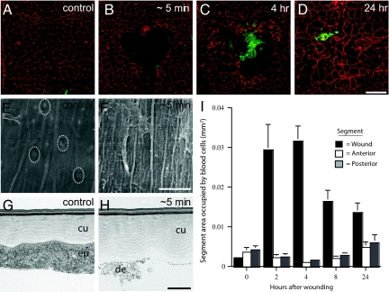
Fig. 2.
Accumulation and turnover of blood cells at larval wounds. (A–D) Unwounded segment (A) or pinch-wounded segments (B–D) in epidermal whole-mount preparations of Pxn>GFP third-instar larvae stained with anti-Fasciclin-III (red) to show epidermal membranes and anti-GFP (green) to show blood cells. (A) Unwounded control larva; (B) ≈5 min after wounding; (C) 4 h after wounding; (D) 24 h after wounding; (E and F) SEM; (E) unwounded epidermis (white dotted ovals, epidermal cell nuclei); (F) exposed cuticle and cell debris ≈5 min after wounding; (G and H) TEM; (G) unwounded epidermis (ep) and apical cuticle (cu); (H) intact cuticle and cell debris (de) ≈5 min after wounding; (I) area of epidermis (+/− SEM) occupied by blood cells in wounded dorsal segments (black bars), adjacent segments located anterior (white bars), and posterior (gray bars). [Scale bar (D) 100 μm for A–D;. (F) 33 μm for F, 26.5 μm for E; (H) 2 μm for G and H.]

