Figure 1.
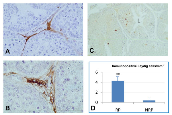
Representative immunohistochemical distribution and localization of D-Asp in the testes of the duck A. platyrhynchos during the reproductive period (RP) (A and B) and the non reproductive period (NRP) (C). D-Asp immunopositivity was mainly localized in Leydig cells of the RP (A and B) and completely absent in the NRP (C). Histograms represent quantification of immunopositive Leydig cells/mm2 (D). **, p < 0.01 RP versus NRP. L, lumen. Scale bars: a, 50 μm; b, 20 μm; c, 100 μm.

