Figure 3.

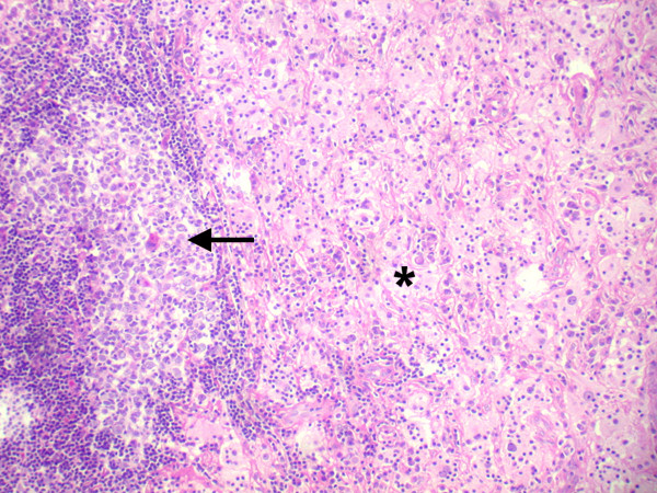
Histopathologic section showing a lymphoid follicle (arrow) surrounded by a cuff of lymphocytes and plasma cells with a few pale areas consisting of sheets of histiocytes (asterisk) (× 50, hematoxylin, eosin).

Histopathologic section showing a lymphoid follicle (arrow) surrounded by a cuff of lymphocytes and plasma cells with a few pale areas consisting of sheets of histiocytes (asterisk) (× 50, hematoxylin, eosin).