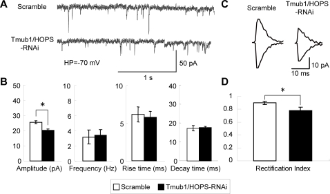
Figure 3. The amplitude of AMPA-mEPSC is suppressed in Tmub1/HOPS-RNAi neurons in a GluR2-containing AMPAR dependent manner.
(A) Representative traces of AMPAR-mediated mEPSC. (B) The AMPA mEPSC amplitude of the Tmub1/HOPS-RNAi neuron was significantly smaller than that of the scramble-treated neuron (n = 10; *P<0.05; t-test). In contrast, the AMPA mEPSC frequency, rise time, and decay time of the Tmub1/HOPS-RNAi neuron did not differ significantly from those of the scramble-treated neuron (n = 10; P>0.05; t-test). (C) Averaged mEPSC at holding potential of +50 (top) and −70 mV (bottom) recorded from the control and HOPS-RNAi induced primary cultured neuron. (D) Rectification index of the HOPS-RNAi induced neuron showed the significant decrease compared to the control neuron (n = 10; *P>0.05; t-test).

