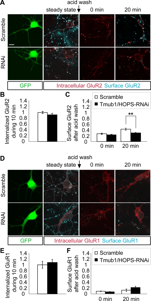
Figure 5. Tmub1/HOPS-RNAi decreases the recycling of internalized GluR2, but not of GluR1, to the cell surface.
(A) The representative images of the GluR2 recycling assay on Tmub1/HOPS-RNAi and scramble-transfected neurons. Live neurons were stained with the anti-GluR2 antibody and were incubated for 10 min for internalization. After the internalization period, the antibodies remaining on the surface were removed using an acid buffer. Then, the neurons were further incubated for returning of the antibody-GluR2 complex to the cell surface. After fixation, the surface-recycled GluR2 was detected with a secondary antibody, and the neurons were then permeabilized followed by the detection of intracellular GluR2 with another secondary antibody. (B) The normalized value of internalized GluR2 during the first 10 min. The internalized GluR2 level did not differ significantly between the Tmub1/HOPS-RNAi- and scramble-transfected neurons (n = 12; P>0.05; t-test). (C) The normalized value of surface GluR2 depending on duration of incubation after the acid wash. After the incubation of 20 min, the recycling of internalized GluR2 to the cell surface was significantly delayed in Tmub1/HOPS-RNAi-transfected neurons as compared to the scramble-transfected neurons (n = 27; **P<0.01; t-test). (D) The representative images of the GluR1 recycling assay on the Tmub1/HOPS-RNAi- and scramble-transfected neurons. (E) The normalized value of internalized GluR1 during the first 10 min. The internalized GluR1 level did not differ significantly between the Tmub1/HOPS-RNAi- and scramble-transfected neurons (n = 9; P>0.05; t-test). (F) The normalized value of surface GluR1 depending on the duration of incubation after the acid wash. After the incubation of 20 min, the recycling of internalized GluR1 did not differ significantly between the Tmub1/HOPS-RNAi-transfected and scramble-transfected neurons (n = 9; P>0.05; t-test). The fluorescence intensity was normalized by the intensity of the internalized AMPARs during the first 10 min in the scramble-transfected neurons. The values shown indicate the means±SEM. Scale bar, 10 µm.

