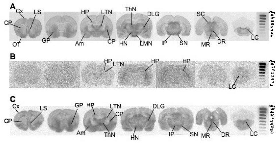
Figure 3.
Ex vivo autoradiograph shows [18F]1 localization in rat brain at 60 min post tracer injection. A: While under isoflurane anesthesia 3.6 mCi was injected through the femoral vein. Tracer localization is seen in regions with medium to high SERT density. B: Escitalopram (2mg/kg), a potent SERT inhibitor, was injected 5 min prior to [18F]1 (4 mCi) injection. Significant inhibition in regional brain uptake was observed in a rat pretreated with escitalopram. The results strongly suggest that [18F]1 and escitalopram compete for the same binding sites in the brain. Low amounts of non-SERT binding is seen in the HP, LTN, and LC. C: Nisoxetine (10 mg/kg), a selective NET inhibitor, was injected 5 min prior to [18F]1 (4.8 mCi) injection. Some inhibition in the locus coeruleus is seen, but otherwise [18F]1 localization is similar to that observed in A. The images suggest that [18F]1 is binding to SERT and not NET. An 125I standard was exposed with each film to allow for a rough comparison between images generated from separate experiments. Cx: cortex; LS: lateral septum; OT: olfactory tubercle; CP: caudate putamen; GP: globus pallidus; HP: hippocampus; LTN: lateral thalamic nuclei; Am: amygdala; ThN: thalamic nuclei; DLG: dorsal lateral geniculate nucleus; LMN: lateral mammillary nuclei; HN: hypothalamic nuclei; IP: interpeduncular nucleus; SN: substantia nigra; SC: superior colliculus; DR: dorsal raphe; MR: medial raphe; LC: locus coeruleus.

