
Figure 3. Real-Time Intraoperative NIR Fluorescent Sentinel Lymph Node Mapping.
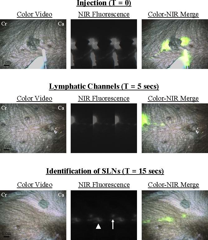
At T = 0, four peri-tumoral, subcutaneous injections of 1 nmol of HSA800 were made around a primary melanoma on the ventral left torso. Two dominant lymphatic channels, one cranial (Cr) and one caudal (Ca) were found. The caudal channel was followed (T = 5 secs) until two SLNs were identified at T = 15 secs. Images shown include color video (left), NIR fluorescence (middle) and a pseudo-colored (lime green) merge of the two (right). Exposure time (67 msec) and normalizations were the same for all NIR fluorescence images.