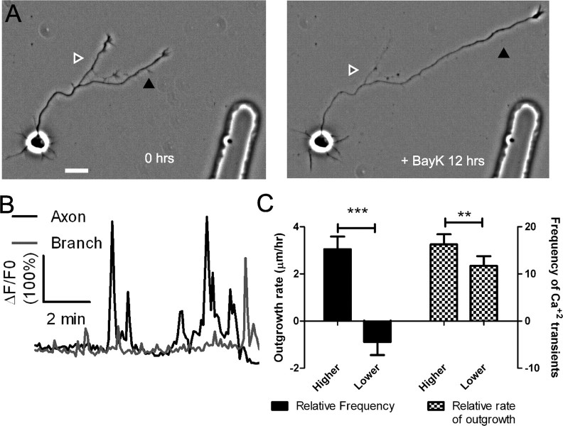
Figure 6.
BayK-induced calcium transients persist throughout long term treatments. A, Phase images of a cortical neuron treated with BayK for 12 h. At the end of the imaging session the processes on right (black arrowhead) extended, whereas the process on the left retracted (white/gray arrowhead). B, After phase imaging, cortical neurons were loaded with Fluo-4 and calcium activity was measured. The extending process from A (black) showed high levels of calcium activity compared with the retracting process (gray). C, Quantifications from 16 experiments imaging calcium activity after long-term BayK treatments revealed correlations between BayK-induced differential calcium activity and outgrowth. C, Left, Comparisons between processes from the same axon showed that processes with higher frequencies of BayK-induced calcium transients had higher rates of outgrowth, whereas processes with lower frequencies retracted (n = 22 comparisons; ***p < 0.001, paired t test). Right, Processes that extended faster had higher frequencies of calcium transients (n = 26 comparisons; **p < 0.01, paired t test). Scale bar, 20 μm.

