Figure 9.
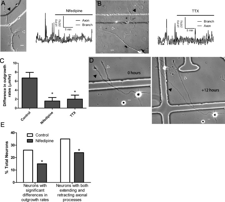
Nifedipine silences localized calcium transients and decreases differential outgrowth. A, DIC image (left) and graph of calcium activity (right) of a neuron with localized calcium transients in the axon (black arrowhead) and the branch (white/gray arrowhead) silenced by nifedipine. Robust localized calcium activity was almost completely silenced after addition of nifedipine (indicated by black bar). Nifedipine decreased localized calcium activity in five of six experiments. B, An example of a neuron in which TTX reduced localized calcium activity. TTX reduced localized calcium activity in five of five experiments. C, Differences in rates of outgrowth among processes from the same axon were smaller in neurons in which localized calcium activity was silenced by nifedipine (n = 5) and TTX (n = 5) than in control neurons (n = 12) (*p < 0.05, ANOVA on ranks with Dunn's posttest). D, An example of process outgrowth during 12 h of nifedipine application. Both processes (white/gray and black arrowheads) showed robust outgrowth at approximately the same rates. E, Bath application of nifedipine in long-term experiments (n ≥ 130 cells from 5 experiments for each condition) decreased the number of neurons with significant differences in the rates of outgrowth of their axonal processes (left) and decreased the number of neurons with both extending and retracting axonal processes (right, *p < 0.05, Fisher's exact test). Scale bars: 10 μm.

