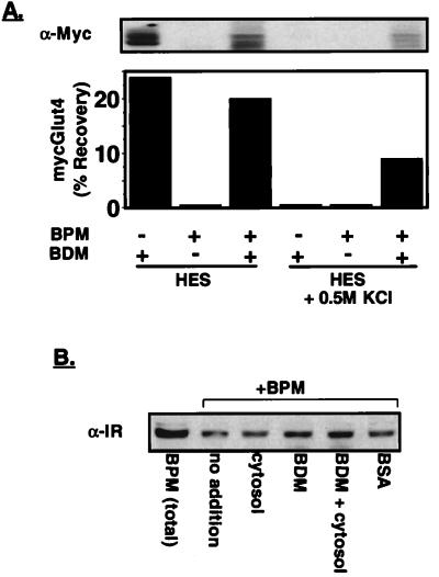
Figure 3.
Optimization of assay conditions I: dilution buffer and the recovery of membrane fractions. The donor membrane was prepared from 3T3-L1 adipocytes expressing mycGlut4. The PM and the cytosol were prepared from nontransfected 3T3-L1 adipocytes. The membrane preparations were incubated for 15 min at 37°C in a total volume of 75 μl [assay buffer included 20 mM Hepes-KOH (pH 7.0), 250 mM sucrose, 0.5 mM EGTA, 1.5 mM MgCl2, 0.5 mM CaCl2, 1 mM DTT, 50 μg/ml BSA, 50 mM KCl, and an ATP-regenerating system]. (A) The assay mixtures were diluted with homogenization buffer without (left three lanes) or with (right three lanes) 0.5 M KCl and then subjected to 15,000 × g centrifugation for the reisolation of the PM. The association between the PM and the intracellular mycGlut4-containing vesicles was analyzed by detection of mycGlut4 in the reisolated PM by anti-myc immunoblot (Upper). The immunoblots were quantified by scanning densitometry, and the data are expressed as a percentage of the total mycGlut4 included in the assay (Lower). (B) To test factors that might influence the recovery of PM, cytosol, BDM, or BSA was included during the incubation period and PM was recovered as described above. The recovery of the PM after reisolation was assessed by the recovery of the insulin receptor by immunoblotting with antibody to the β-subunit of the insulin receptor.

