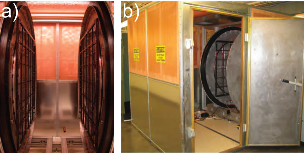
Figure 1.

Photographs of the open-access human MRI system. a) The open-access imaging area, which allows reorientation of a subject. The gap between the two coils is 90 cm, with over 2 m of open space in the other two dimensions. The photograph shows the pair of main B0 coils on their aluminum support flanges, with the gradient coils located parallel to each B0 coil on additional supports bolted to the flanges. b) The entire imager on its customized aluminum framework, located inside an RF-shielded room. Access to the imaging region from outside the room is straightforward.
