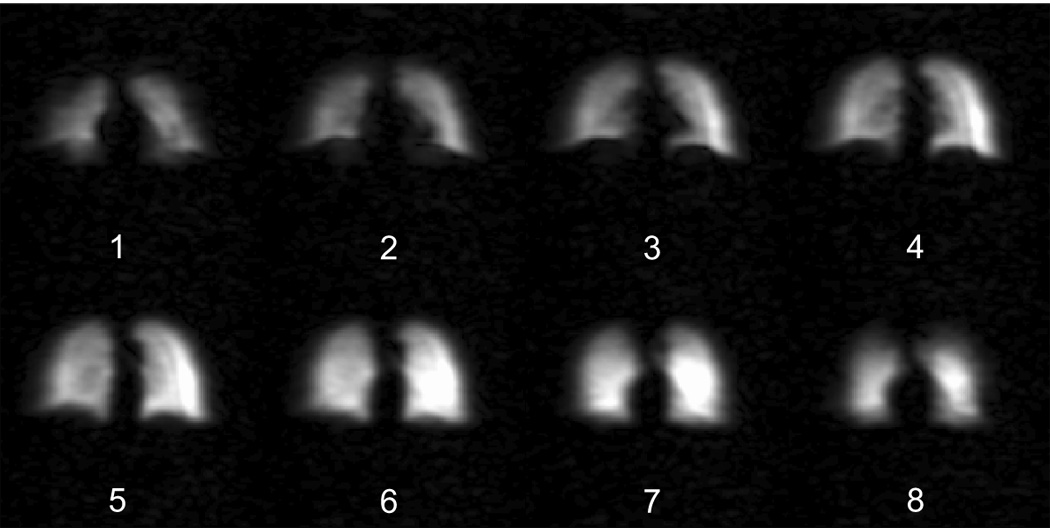
Figure 4.

Three-dimensional 3He MR image series of human lungs, obtained using the open-access human MRI system, with subject lying horizontally in a supine orientation. All planes visualize the lungs as if looking at the subject from the front – i.e., the subject’s right lung lobe is on the left of the image. Image planes represent slices ~ 1.5 cm thick, and progress from anterior (# 1) to posterior (# 8) through that dataset. Imaging parameters: B0 = 6.5 mT, Larmor frequency = 210 kHz, FOV = 50 × 50 × 12 cm, NEX = 1, flip angle = 4°, TE/TR = 28.5/85.8 ms. Data size = 128 × 64 × 6, zero-filled to 128 × 128 × 8, total scan time ~ 30 s.
