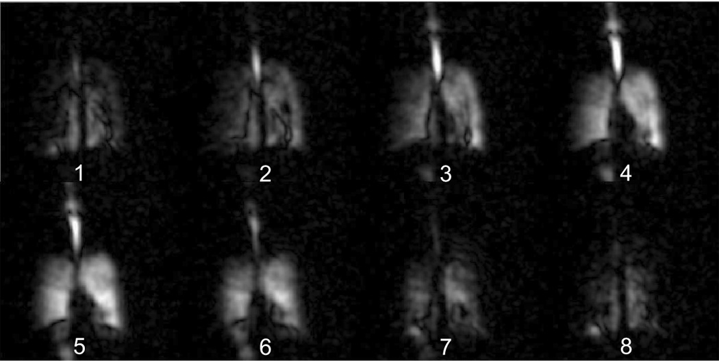
Figure 5.

Three-dimensional 3He MR image series of human lungs, obtained using the open-access human MRI system, with subject positioned vertically. Additional room air was not inhaled following 3He inhalation, resulting in non-uniform 3He distribution throughout the lung, and intense signal in the trachea and oral cavity. MR signal below the diaphragm in each image, beside the plane number, is most likely due to gas above the trachea and outside the top of the image field-of-view that was folded in to the bottom portion of the image. Image orientation, layout and acquisition parameters are the same as for Figure 4.
