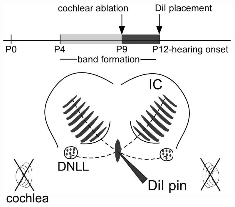
Figure 1.
Schematic illustration of the experimental design. A time line shows the relationship of time points for bilateral cochlear ablation (indicated by crossed out spiral symbols) and DiI labeling of DNLL projections in the IC to the time course of development of afferent bands in the central nucleus of the IC (CNIC) and hearing onset. The line drawing of the dorsal part of the midbrain indicates where DiI pins were positioned to label DNLL fibers as they cross the midline in the dorsal tegmental commissure (of Probst) and distribute in a pattern of afferent bands (shaded regions) in the contralateral CNIC. Other abbreviations: DCIC, dorsal cortex of the IC; ECIC external cortex of the IC.

