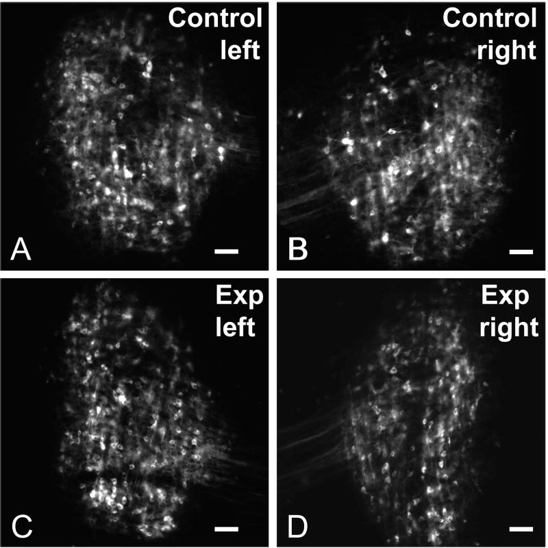
Figure 2.
Paired images illustrating retrogradely labeled neurons in the DNLL for the control group (A, B) and experimental (Exp) group (C, D) cases after DiI-pins were placed in the dorsal tegmental commissure (see Experimental Design in Figure 1) to label decussating fibers. Note the symmetric labeling in DNLL on each side in both groups. Scale bars equal 50 μm.

