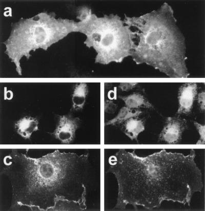
Figure 3.
Subcellular localization of HMW 4.1R isoforms. COS-7 cells transfected with each of the cloned HMW 4.1R cDNAs were labeled with different antibodies and examined by epifluorescence microscopy 48 h after transfection. (a) Cells stained with anti-c-Myc antibody; (b and d) cells double stained with anti-c-Myc antibody (b) and anti-PDI antibody (d), a marker of the ER; (c and e) cells double stained with anti-4.1R (10b) antibody (c) or anti-CD4 mouse monoclonal antibody (e), a marker of the plasma membrane. Representative images are shown: (a) predominant expression at the plasma membrane and the ER; (b) at the ER; (c) at the plasma membrane. These expression patterns were observed for the six HMW 4.1R isoforms.

