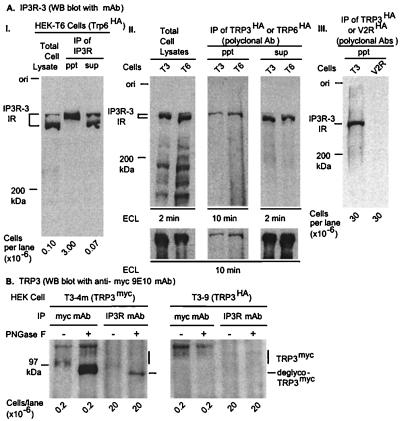
Figure 1.
Reciprocal coimmunoprecipitation of IP3R and TRP. T3–9 or T6–12 cells, stably expressing HA epitope-tagged TRP3 or TRP6, were lysed, and immunoreactive IP3R was analyzed by Western blotting. (A) IPs of TRP3 and TRP6 contain type-3 IP3R. (I) IP3R-3 in cell lysates, immunoprecipitates (IP, ppt) and IP supernatant (IP, sup). (II) IP3R-3 in detergent extracts and in the respective IP ppt and IP sup. Lower panels of A.II provide an idea of the relative proportion of total IP3R coimmunoprecipitated with TRP3 or TRP6. (III) Lack of significant nonspecific coimmunoprecipitation of IP3R3 immunoreactive material in an IP of V2R, an unrelated integral membrane protein expressed stably in HEK cells (32). Western blot with anti-human IP3R-3 mAb. (B) IPs of IP3R from T3 cell extracts contain TRP3. Detergent extracts of HEK-T3 cells stably expressing TRP6myc or TRP3HA were treated or not with PNGase-F and incubated overnight either with anti-type-1/2/3 IP3R mAb or with 9E10 anti-myc mAb, both preadsorbed to protein-A Sepharose. IPs were washed, dissolved, and electrophoresed as described in Materials and Methods. TRP3myc in IP3R and TRP3 IPs was visualized with 9E10 mAb and detected by enhanced chemiluminescence (ECL).

