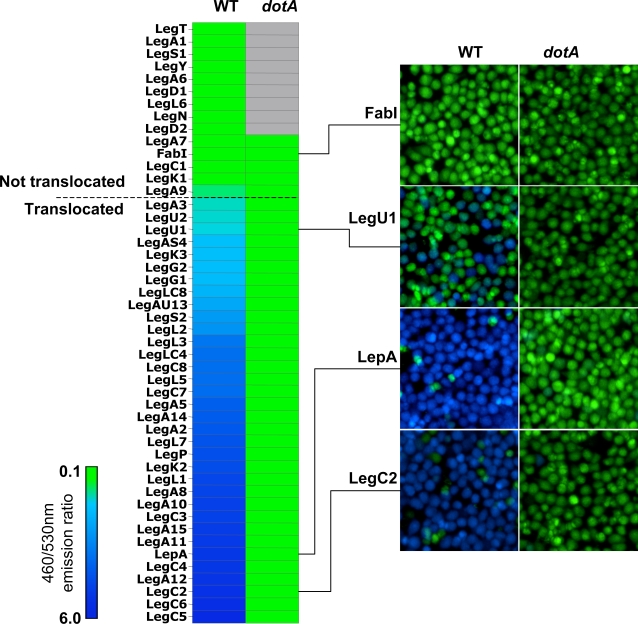
Figure 1. Icm-Dot Dependent Translocation Leg Substrates into J774 Cells.
J774 cells were infected with KS79 (WT) or KS79dotA harboring TEM1-Leg protein fusions at an MOI of 50. Infected cells were loaded with CCF4/AM and translocation was determined by measuring the ratio of cleaved (460 nm) to uncleaved (530 nm) CCF4/AM. Ratios of 460/530 nm emission for each TEM1-Leg protein in wild type KS79 (WT) or KS79 dotA is shown as a heat diagram. A 460/530 nm ratio of more than 1 indicates translocation and is indicated by a horizontal dashed line. Representative images obtained using epifluorescence microscopy on individual assay wells is shown on the right. The results shown represent the average of 2 to 3 experiments, each performed in triplicate.

