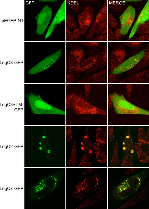
Figure 7. Localization of Leg-GFP Fusion Proteins and KDEL-containing proteins in transiently transfected CHO-FcγRII cells.
CHO-FcγRII cells were transfected with plasmids expressing GFP (pEGFP-N1), LegC3-GFP (pRG6), LegC3ΔTM (pRG7), LegC2-GFP (pRG8) or LegC7 (pRG10), as indicated on the left of each row. Representative confocal images were acquired demonstrating the location of GFP and proteins containing KDEL motifs as determined by immuno-fluorescent staining. Merged images are shown in the right column where yellow color designates overlap of the green and red channels.

