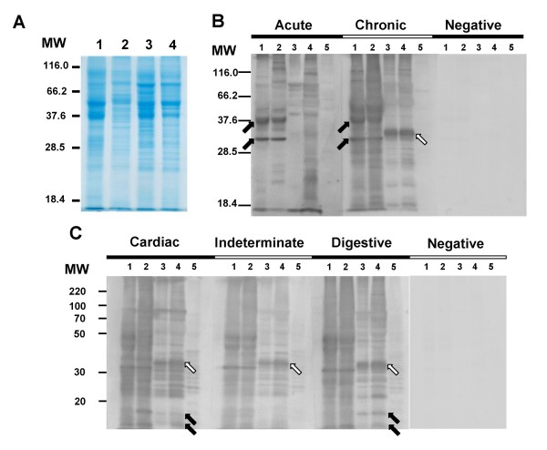
Figure 3.
Immunoblot profiles of Trypanosoma cruzi (Y strain) and Trypanosoma rangeli (Choachi strain) with chagasic sera. Total protein extracts resolved in a 12% SDS-PAGE and stained by Comassie brilliant blue (A) and the immunoblot analysis of the same lysates using sera from acute and chronic chagasic patients (B) or using sera from chronic chagasic patients with the cardiac, indeterminate or digestive forms of the disease (C). On panel B, arrows indicates proteins recognized by chagasic sera (acute and/or chronic) on T. cruzi (dark arrows) or on T. rangeli (white arrow) extracts. Arrows in panel C indicates a T. rangeli 11 and 15 kDa proteins exclusively recognized by the cardiac and digestive serum in both T. rangeli forms (dark arrows) and a 35 kDa protein (white arrows) recognized by all sera from T. cruzi-infected patients. Lanes 1 and 2 = T. cruzi Y strain epimastigote and trypomastigote forms and lanes 3 and 4 = T. rangeli Choachi strain epimastigote and trypomastigote forms, respectively. Lane 5 = Vero cells extract; MW = molecular weight marker (kDa). Asterisks indicates significant differences (p < 0.05).

