
Figure 3. Electrical stimulation evokes nitric oxide production in the mouse olfactory bulb slice.
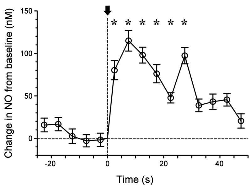
An NO micro-sensor was inserted into the granule cell layer of a slice (depth ~ 50 μm) and the electrode current was recorded as the slice was stimulated by current pulses from concentric bipolar electrode placed in olfactory nerve layer radial to the sensor site (a 200 ms train of 20 current pulses at 100 Hz, initiated at time 0, indicated by arrow). The plot shows data sampled at 20 Hz binned in 5 s intervals (mean values with standard deviations indicated). Starred values are significantly greater than mean pre-shock values (p < 0.05, Mann Whitney rank sum test). Electrode calibration was 24 nM/pA.