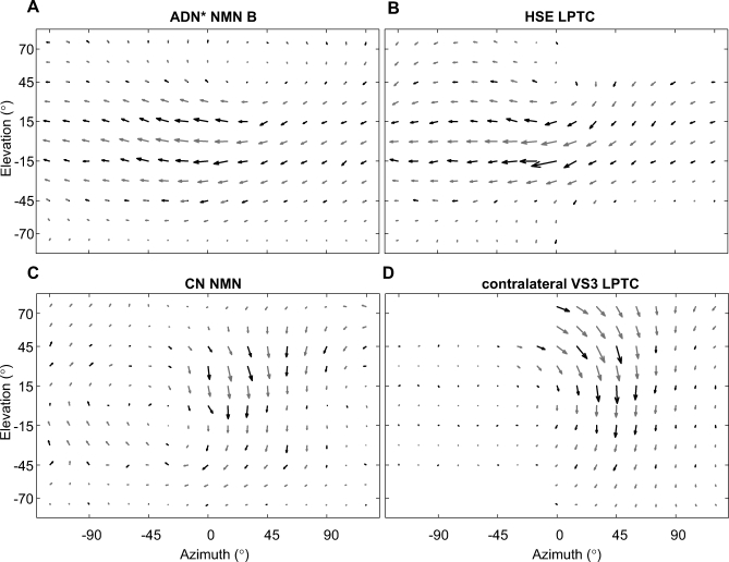
Figure 5. Comparison of NMN and LPTC Receptive Field Maps.
(A) The receptive field of an ADN* NMN, obtained using the grating stimulus. The recording was made from the TH muscle group. The known NMN-muscle connectivity [25] strongly suggests that this unit belongs to the ADN. The asterisk indicates that this nerve assignment is based on anatomical criteria. The receptive field of this NMN is similar to that of an identified HSE tangential cell shown in (B). Both the ADN NMN and the HSE LPTC have receptive fields reminiscent of the optic flow field induced by yaw rotation. HSE data were taken from [29].
(C) The receptive field obtained from a NMN in the left CN, using the dot stimulus, is similar to that of the contralateral identified VS3 tangential cell shown in (D). Both the CN and VS3 receptive fields are similar to an optic flow field generated during nose-up banked turn to the left (for further explanation see text). VS3 data were taken from [27].

