
Fig. 1.
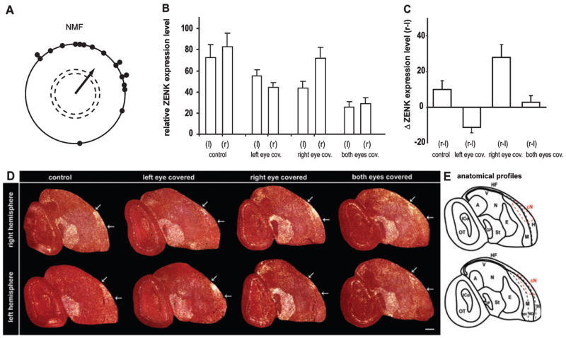
ZENK expression in European robin brains under different eye-cover conditions. (A) Orientation of European robins during spring migration in an NMF in our behavioural apparatus. Each dot represents the mean orientation of one individual bird. The arrow indicates the mean orientation of the group. The length of the arrow represents the length (r-value) of the group mean vector. The dashed circles indicate the radius of the group mean vector needed for significance (P < 0.05 and P < 0.01) according to the Rayleigh Test of uniformity. (B) ZENK expression levels in Cluster N of the right (r) and left (l) hemisphere for four groups of birds with various eye open and covered conditions. (C) Direct within-bird subtracted comparisons of the interhemispheric differences in ZENK expression levels for the four experimental groups. One-way ANOVA comparison between all groups revealed highly significant differences for all group comparisons (P < 0.001), except for the comparison between the two control groups: birds with both eyes open (control) and birds with both eyes covered. (D) Darkfield image of ZENK expression in right and left hemisphere saggital sections showing Cluster N expression under different eye-cover conditions. White arrows mark the caudal and rostral boundaries of Cluster N for each brain section. Rostral is right, dorsal is up. (E) Anatomical profile of the right image (condition: both eyes covered) shown in (D). Abbreviations: A, arcopallium; Cn, Cluster N; E, entopallium; GP, globus pallidus; HF, hippocampal formation; H, hyperpallium; ICo, inferior colliculus; M, mesopallium including ventral (MV) and dorsal (MD) part; N, nidopallium; OT, optic tectum; St, striatum; v, ventricle; W, visual Wulst. Error bars are SEM. Scale bar, 1 mm.