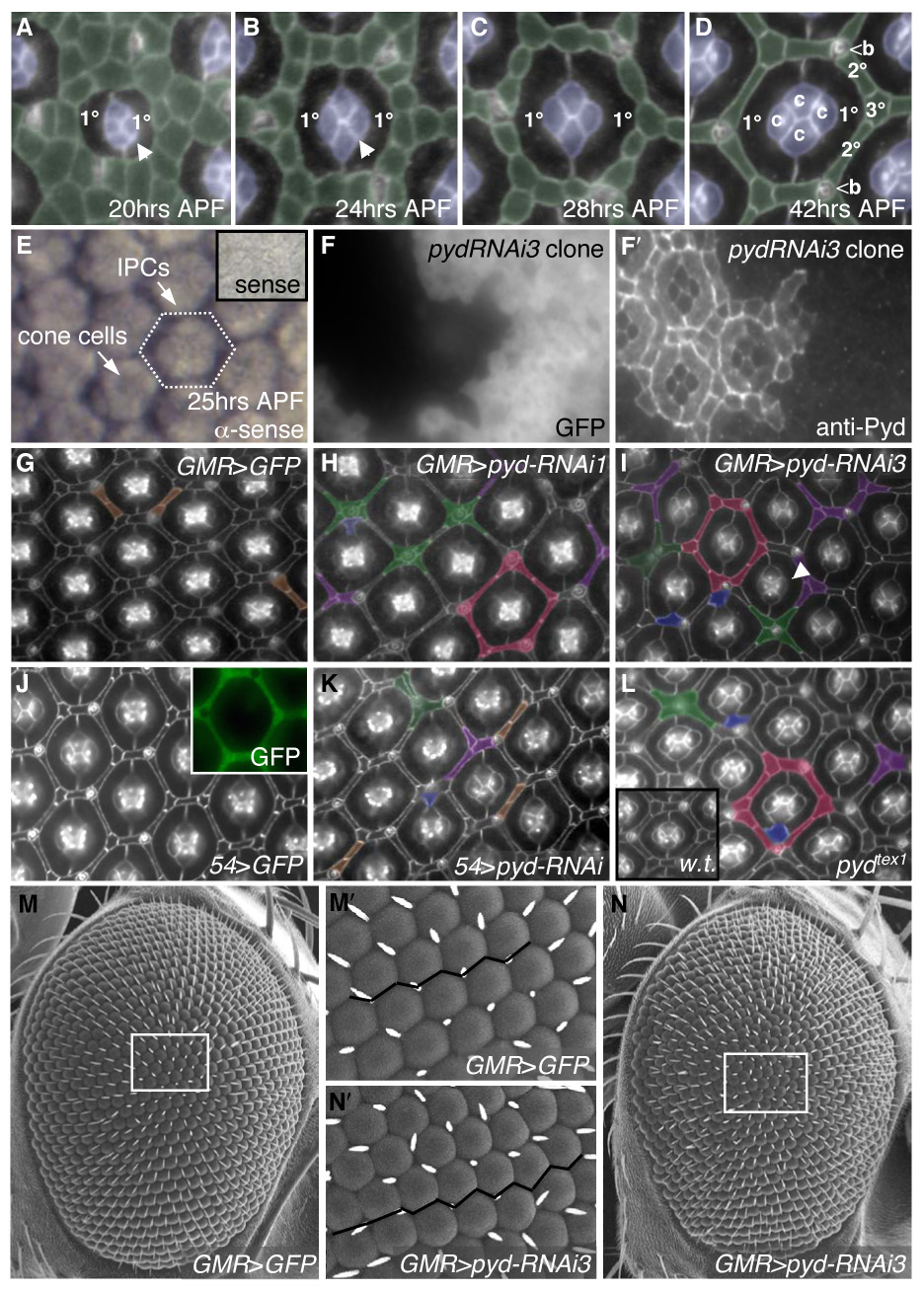
Fig 1.

pyd regulates patterning of IPCs. Apical cell profiles were detected with anti-Arm antibodies at 41–42 hours APF (after puparium formation) unless otherwise indicated. Anterior is to the right in this and all subsequent images. A–D. Normal development of the pupal eye is marked by sorting of the IPCs (pseudo-colored in green) into a hexagonal lattice. Between 18–24 hours APF, cell intercalation narrows the rows of IPCs to single file (B). The hexagon is composed of six 2°s forming the sides of the hexagon and three 3°s at the vertices, alternating with bristles (b) (D). E–E′. In situ hybridization using full-length pyd-RB anti-sense probe in 25 hr APF pupal retinas: pyd was expressed at highest levels in IPCs and at lower levels in cone cells and 1°s. Inset shows pyd-RB sense probe. F–F′. Expression of pyd-RNAi3 in clonal patches in the eye marked by GFP (F) demonstrated cell-autonomous loss of Pyd antibody staining at the apical membrane (F′). G–L. Examples of patterning defects are pseudo-colored as follows: extra cells in the absence of other defects (brown), clustering of cells around bristles (green), pentagonal ommatidia (red), IPC-cone contact cells (blue) and a failure to specify a single 3° cell (purple). G. Control GMR>GFP retinas. H–I. GMR>pyd-RNAi1 (H) or GMR>pyd-RNAi3 (I) resulted in far stronger IPC patterning defects. Note also the failure to switch cone-cell contacts (I; arrowhead). J. 54>GFP retinas have no patterning defects (J). Expression of 54-Gal4, visualized with GFP, is found primarily in IPCs (inset). K. 54>pydRNAi exhibited errors in IPC organization. L. pydtex1 mutant eyes had defects that were similar to RNAi expression; inset shows wild-type retina. M–N. SEM of control (M) and pyd-RNAi3 (N) expressing eyes. The white tracing outlines an area that is shown at higher magnification in M′ and N′, respectively. The black line emphasizes the slightly uneven ommatidial rows in pyd-RNAi expressing eyes (N′) vs. control (M′).
