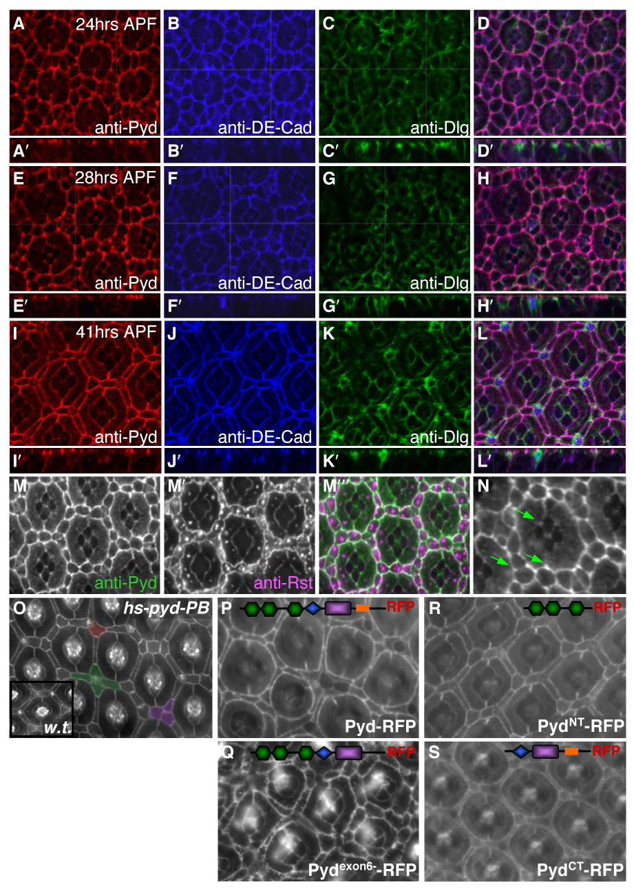
Fig 2.

Pyd localized to AJs and its over-expression led to pupal eye patterning defects. A–L. Apical confocal sections of wild type pupal eye were taken at 24 hours APF (A–D), 28 hours APF (E–H) and 41 hours APF (I–L). Anti-Pyd immunofluorescence (A, E, I) co-localized extensively with anti-DE-Cad (B, F, J) at the AJ but only overlapped slightly with the septate junction marker Dlg (C, G, K). D, H, L. Overlay of anti-Pyd, anti-DE-Cad and anti-Dlg. Inset panels A′–L′ are lateral projections. M–M″. Anti-Pyd (M) co-localized with anti-Rst (M′) at the IPC/1° interface at 28 hours APF (overlay in M″). N. Anti-Pyd was concentrated where three or more cells came together (‘tricellular junctions’; green arrows). O. 41 hours APF pupal CS (inset) or hs-pyd-RB (O) eyes with a 30 min 37°C heat-shock at 17 hours APF. Notice that some phenotypes were shared with pyd-RNAi, such as the clustering of cells around bristles (pseudo-colored green) and the failure to specify a single 3° cell (pseudo-colored purple). Occasionally, a 2° extended to cover both the 2° and 3° niches (pseudo-colored red). P–S. GMR>UAS-pyd-RFP expression in 42 hr APF eyes; the relevant domains of each construct are schematized (see Supp Fig 3A). Pyd-RFP (P) localized to the AJ and directed patterning defects similar to those observed with pyd-RNAi expression. Pydexon6--RFP localized to the AJ and had severe cone cell and IPC defects (Q). PydNT-RFP and PydCT-RFP localized to the AJ but both rarely exhibited patterning defects (R and S, respectively).
