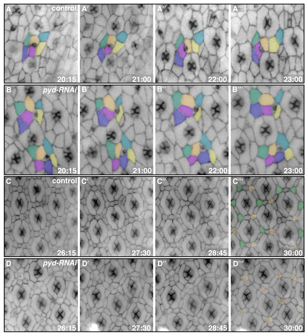
Fig 3.

pyd-RNAi expressing cells failed to execute productive movements or sort appropriately. Membranes were labeled with α-Catenin-GFP. Times represent hours APF. A–B‴. Panels from four time points during cell intercalation for both control (A–A‴) and pyd-RNAi (B–B‴) retinas. Cells were pseudo-colored to emphasize particular cell movements. Typically, pyd-RNAi-expressing cells failed to undergo intercalation, instead remaining in double rows. C–D‴. Panels from four time points during 3° cell patterning for both control (C–C‴) and pyd-RNAi (D–D‴) retinas. Control cells moved into and out of the 3° position early; by 30 hours, however, one cell had taken over the niche in most cases (green in C‴; bristles were pseudo-colored in orange in C‴ and D‴). By contrast, the pyd-RNAi-expressing cells maintained initial contacts and, in most cases, failed to establish a single 3° (D‴).
