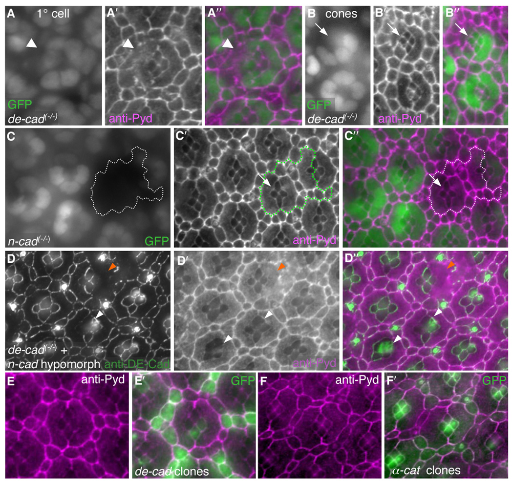
Fig 7.

DE-Cad is necessary for Pyd to localize to the AJ. All images taken at 28 hours APF. A–B′. Single-cell null de-cad/shotgun (shg1H) 1° clones (A, arrowhead) and cone cell clones (B, arrow) are marked by loss of nuclear GFP expression. In isolated 1° cell clones (A), Pyd de-localized from the membranes of the adjoining IPC and cone cells that contacted the de-cad deficient cell membrane (A′, arrowhead). Pyd was maintained at the cone/cone interface in de-cad null cone cell clones (B′, arrow). C–C″. Null n-cadherin (n-cadM19) clones, marked by lack of nuclear GFP (C), showed no change in Pyd localization (C′) at the cone/cone interface (arrow, overlay in C″). D–D″. Null de-cad (shgR69) single-cell clones (marked by loss of DE-Cad staining in D) in the background of a strong reduction in n-cadherin (n-cadM12/n-cadM19) exhibited de-localized Pyd staining at the cone/cone interface (D′, white arrowheads). Null de-cad clones in 1°s were also observed (orange arrrowheads). D″ shows overlay. E–E″. DE-Cad-over-expression in single cells (labeled with GFP, green in E′) did not alter the localization or level of anti-Pyd immunofluorescence (E and magenta in E′). F–F′. UAS-α-catenin expression in single cells (labeled with GFP, F′) did not alter the localization or level of anti-Pyd immunofluorescence (F and magenta in F′).
