Fig 8.

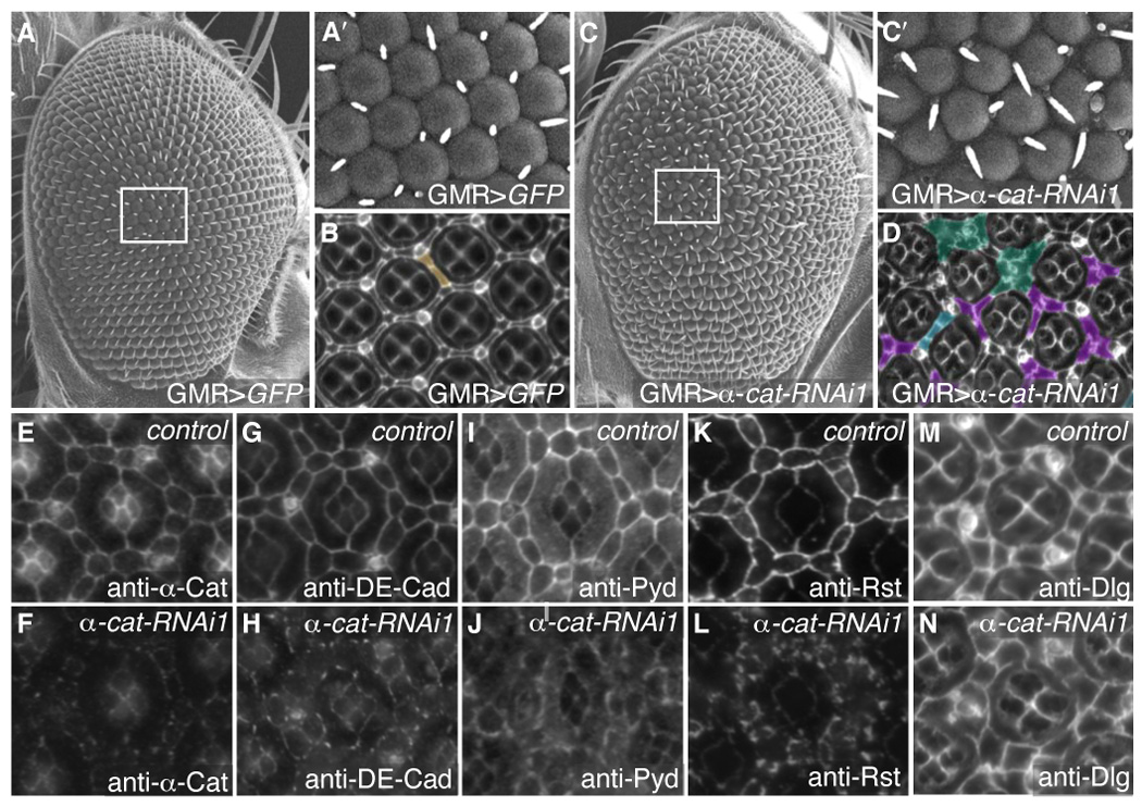
α-Catenin is necessary for AJ protein localization. A–A′. SEM of control GMR>GFP adult eyes demonstrating even ommatidial rows (A) with boxed area shown enlarged in A′. B. 41 hours APF pupal eye stained with anti-Discs large to mark cell membranes. Extra cell was pseudo-colored in brown. C–C′. Reduction of α-Catenin (GMR>α-cat-RNAi) disrupted the orderly array of ommatidial rows in the adult eye (C). C′ shows an enlargement of the boxed area C. D. 41 hours APF pupal eye stained with anti-Discs large and pseuso-colored to show examples of patterning errors: extra cells (blue), clustering of IPCs around bristles (green), and the failure of only one cell to occupy the 3° niche (purple). E–N. 28 hours APF pupal eyes. E–F. GMR>α-cat-RNAi retinas showed greatly reduced levels of α-Catenin (F) compared to GMR>GFP expressing eyes (E): the image in F represents >8-fold longer exposure than E. G–N. Control GMR>GFP retinas visualizing DE-Cad (G), Pyd (I), Rst (K) and Dlg (M). Expression of GMR>α-cat-RNAi in the pupal retina resulted in de-localization of DE-Cad (H), Pyd (J) and Rst (L), but no change in anti-Dlg staining (N).
