Figure 1.
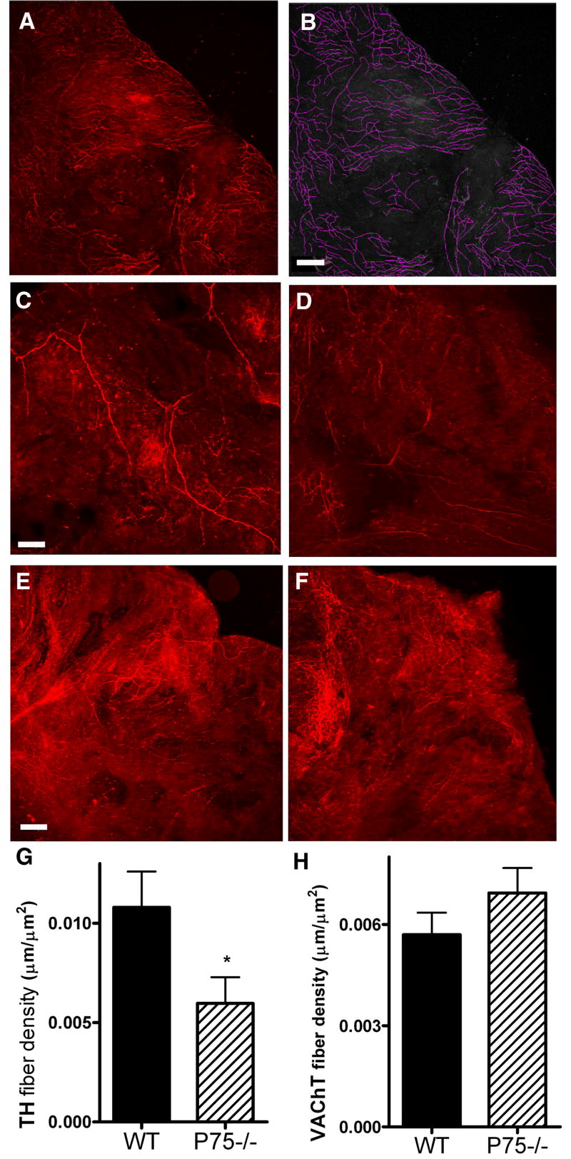
Sympathetic, but not parasympathetic innervation of the right atria is altered in P4 p75 knockout mice. Right atria were stained for TH to identify sympathetic fibers or VAChT to identify parasympathetic fibers. Confocal images were captured at 10 μm intervals and fibers were manually traced using from a compressed z-stack using ImageJ software with the NeuronJ plug-in. Fiber density was calculated for each atria. Panels A and B show an example of fiber tracing on an image taken from an adult atrium stained with VAChT. C, D: Representative TH staining from P4 WT (C) and p75-/- (D) atria. E, F: Representative VAChT staining from P4 WT (E) and p75-/- (F) atria. Images C-F show representative images from the same region of the right atria, fiber tracing was carried out over the entire atrium. G, H: Average TH (G) and VAChT (H) fiber densities for wildtype (WT, solid bars) or p75-/- (hatched bars) atria. Data shown are the mean±sem, *p < 0.05, n=10-11 for TH, n=6 for VAChT. Scale bar = 125 μm.

